A-Scan
Features
Biometry/Pachymetry modes: Auto Manual Super Auto.
Lightweight Desktop Design.
Easy To Use Interface.
Built In Printer(Full Report, Current Screen).
Computer Ready Interface(Compact Flash, USB, Internet).
Patient Records: Excel or binary format.
Improved Contact and Immersion Capability.
LASIK Pachymetry option.
K Adjusment Software for Post-Refractive Cataract Screening.
Built-in Contact Eye Model.
Large SVGA Color LCD/Touch Screen.
Age Compensation Option.
Adjustable Screen Brightness.
White To White Input for Patients Records.
Velocity Programmable for each Segment.
IOL Constants Customization Software.
Allows up to 5 Users Each in Biometry and Pachymetry to Store Individual System setting.
Language option: Deutsch, English, Espanol, Francis,Italiano,Japanese, Portuguese.
Alcon Realibility and services.
AL-Scan
10 seconds to measure 6 values
In 10 seconds, six values for cataract surgery are measured:
• Axial length
• Corneal curvature radius
• Anterior chamber depth
• Central corneal thickness
• White-to-white distance
• Pupil size
3-D auto tracking and auto shot
The AL-Scan incorporates NIDEK’s much acclaimed 3-D auto tracking and auto shot, which provides the operator with the most ease, comfort, and accuracy on all measurements
Ability to measure eyes with even dense cataract
Advanced measurement algorithms enhance the signal-to-noise ratio, which allows the AL-Scan to measure eyes with even dense cataract.
Optional built-in ultrasound biometer
In cases where the optical biometer cannot measure an eye with an extremely dense cataract, the AL-Scan provides an optional built-in ultrasound biometer, allowing measurement of virtually any cataractous eye.
Anterior segment observation with imaging of lens, pupil, and double mire rings
The AL-Scan provides sectional lens image, pupil image, and reflected image of double mire rings, which enables the operator to observe the anterior segment.
IOL calculation with its own measured values
Nine IOL calculation formulas are incorporated in the AL-Scan. Once measurement is completed, the IOL power is automatically calculated using its own measured data.
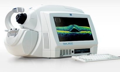
OCT
Optical coherence tomography (OCT) is an optical signal acquisition and processing method. It captures micrometer-resolution, three-dimensional images from within optical scattering media (e.g., biological tissue). Optical coherence tomography is an interferometric technique, typically employing near-infrared light. The use of relatively long wavelength light allows it to penetrate into the scattering medium. Confocal microscopy, another optical technique, typically penetrates less deeply into the sample but with higher resolution. Depending on the properties of the light source (superluminescent diodes, ultrashort pulsed lasers and supercontinuum lasers have been employed), optical coherence tomography has achieved sub-micrometer resolution (with very wide-spectrum sources emitting over a ~100 nm wavelength range). Optical coherence tomography is one of a class of optical tomographic techniques. A relatively recent implementation of optical coherence tomography, frequency-domain optical coherence tomography, provides advantages in signal-to-noise ratio, permitting faster signal acquisition. Commercially available optical coherence tomography systems are employed in diverse applications, including art conservation and diagnostic medicine, notably in ophthalmology where it can be used to obtain detailed images from within the retina.
Selected Applications
Optical coherence tomography is an established medical imaging technique. It is widely used, for example, to obtain high-resolution images of the anterior segment of the eye and the retina, which can, for example, provide a straightforward method of assessing axonal integrity in multiple sclerosis, as well as macular degeneration. Research indicates that OCT may be a reliable tool for monitoring the progression of glaucoma.
OPD Scan 3
Features :
Five-in-One true refractive workstation for all practitioners
The OPD-Scan lll is the Five-in-One true refractive workstation combining
• Wavefront Aberrometer
• Topographer
• Auto Refractometer
• Auto Keratometer
• Pupillometer and Pupillographer
Overview summary for optimal clinical decisions
The overview summary provides essentials of measurement and analysis data for cataract and refractive surgery.
1. Irregularity helps determine the best strategy for vision correction. Separation into Total, Corneal and Internal components allows determination of the source of the optical pathology.
2. PSF images of OPD, Axial, and Internal OPD map simulate objective retinal visual quality from each component of the eye for easy clinical assessment and patient education.
3. Corneal Spherical Aberration aids in the selection of aspheric IOLs and contact lenses.
4. Color coded Classification Indices help identify post-LASIK corneas and Keratoconus.
5. The Astigmatism index aids the implantation of toric IOLs such as incision placement and lens alignment.
6. A retroillumination image of cataracts captured during the OPD exam allows better understanding of pupillary effects on vision and in patient education.
A number of summaries
Besides the overview summary a number of summaries specialized for each clinical case are available.
Wider measurement area
9.5 mm diameter measurement area ensures full coverage of almost any pupil and provides 2,520data points for wavefront aberrometry.
Greater topography resolution, blue placido rings
33 blue placido mires provide a minimum of 11,880 data points. The blue wavelength allows greater precision in ring detection..
Pentacam
If you are thinking of undergoing refractive laser surgery, your doctor will use a Pentacam to accurately measure the thickness, contour, and shape of your cornea. The Pentacam uses a rotating camera to image the anterior segment of the eye. The rotating camera, called a Scheimpflug camera, creates a 3D image. The 3D image is an important topographic map used in LASIK, cataract, and glaucoma surgery. It will also be able to tell your eye doctor if you are a candidate for refractive surgery by screening for keratoconus, a degenerative disorder of the eye in which the cornea’s shape is conical. This causes multiple images, light sensitivity, and streaking.
Different Uses of the Pentacam
By measuring the thickness of the cornea at the back and front, the Pentacam can generate 25,000 data points. This large number of points collected on your eye is an extremely accurate way to detail your cornea’s thickness.
The Pentacam can analyze patients to determine if they would be better candidates for intraocular lens implants than simply laser vision correction. In this way, your ophthalmologist will be able to explain to you exactly why you should get IOLs. With a detailed map to look at, the ophthalmologist can help you to better understand what is going on in your eyes.
The Pentacam is yet another high tech way to make sure the ophthalmologist has the most detailed information he or she can have before your procedure.
To find out more about the Pentacam or any other technological wonders used to map your eyes, please contact an experienced ophthalmologist in your area.
Specular Microscope
Paracentral specular microscopy
In addition to conventional central and peripheral specular microscopy, the CEM-530 includes a unique function to capture paracentral images.The OPD-Scan lll is the Five-in-One true refractive workstation combining
Two-second auto analysis
Once the image is selected, complete analysis is automatically performed in two seconds with the CEM-530.
Auto indication of the optimal image
16 images are captured and automatically sorted based on quality and the ability to be analyzed. The optimal image for analysis is indicated with orange highlight.
Instant printout with built-in printer
The built-in printer provides an instant printout of the analyzed data and images of the
